Fully funded team
Creating virtual reality maps of tumours
 A team of 15 investigators
 UK, Switzerland, USA, Canada and Republic of Ireland
 Biologists, mathematicians, bioinformaticians, astronomers, physicists, clinicians, chemists, neuroscientists
 £20M
£20M
![]() 6 years
6 years
 Find a way of mapping tumours at the molecular and cellular level

Building a tumour in virtual reality allows us to see information about the behaviour, location and characteristics of tumour cells all at the same time. This will help us understand more about tumours and begin to answer questions that have eluded cancer scientists for many years.
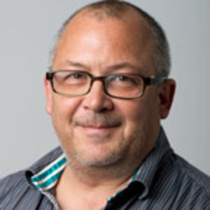
Professor Greg Hannon, Principal Investigator, IMAXT
Background
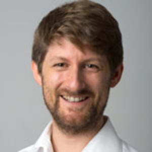
Professor Greg Hannon and the Cancer Grand Challenges IMAXT team were awarded £20 million in 2017 to build 3D tumours, which can be studied using virtual reality.
To fully understand cancer, scientists need to know everything about a tumour – what types of cells are in it, how many there are and where they are located in the tumour.
Having such a detailed picture of a tumour would allow scientists and doctors to develop new ways to diagnose and treat the disease, and new ways to stop it spreading and coming back.
But getting such an accurate, precise picture of tumours is extremely difficult to do. So difficult that it’s not been done before.
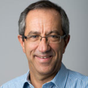
Professor Hannon and his team of scientists, computer scientists and virtual reality experts from England, Canada, Switzerland and the US aim to change this.
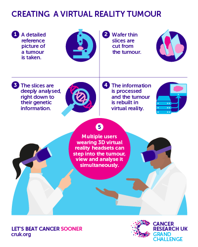
Their Cancer Grand Challenges project aims to build a 3D tumour that can be studied using virtual reality and which shows every single different type of cell in the tumour.
The research
Combining established techniques, including DNA sequencing and imaging, with new technology they will invent and develop, the team will study high-quality breast cancer samples available to them which were collected from women involved in the METABRIC study.
They aim to gather thousands of bits of information about every single cell in a tumour – from cancer cells to immune cells – to find out what cells are next to each other, how they interact with and influence each other, and how they all work together to help tumours survive and grow.
They will then take all the information they collect about the cells in a tumour and use it to construct a 3D version that can be studied using virtual reality.
Using virtual reality will allow scientists to immerse themselves in a tumour, meaning they can study patterns and other characteristics within it, in entirely new ways that aren’t possible in 2D. It will also allow multiple doctors and scientists to look at a tumour at the same time, meaning people at opposite ends of the country, and with different areas of expertise, can work together to help diagnose and treat patients better.
The impact
By developing an entirely new way to study breast cancer, this team hope to change how the disease is diagnosed, treated and managed.
Ultimately, it could improve how women with breast and other types of cancer are classified, which would improve their treatment and help more people survive the disease.
Project update - Year one

The team has already made substantial and promising advances since they started work in May 2017. At the CRUK Cambridge Institute, they have established a central laboratory that will house the cutting-edge imaging equipment and technologies the team is using and developing, and act as the main project hub for sample analysis.
In the first year, the team was committed to getting the VR technology up and running, recruiting a team of VR developers, including programmers and 3D artists, to work alongside the core team. This partnership has allowed experts from different fields to work closely together to design a system that is both state of the art and easy to use by scientists across the world.
The team has successfully produced the first 3D proof-of-principle model of early breast cancer, which was plugged into the VR headset and viewed by multiple users at the same time – a significant milestone. The team will now continue to improve their sample analysis in preparation for uploading real patient data into the VR technology for the very first time.
 Fully funded team